
Tissue Saturation Index (TSI) - Absolute oxygenation measure in local tissues
In this blog, we will discuss Tissue Saturation Index (TSI) as one of the key features used by researchers to answer their research questions. The TSI is a measure of the local oxygenation in the tissue beneath the sensor. It is an absolute value, and it is also known as regional saturation (rSO2) or tissue saturation (StO2).
Background
Hemoglobin, the red blood cells carrying oxygen in blood, absorbs near-infrared light. Oxygenated hemoglobin and deoxygenated hemoglobin have different absorption spectra for near-infrared light. By transmitting near-infrared light into the tissue and measuring the reflected light intensity, relative concentration changes of oxygenated and deoxygenated hemoglobin can be determined via the modified Lambert-Beer law (MLBL).
To obtain absolute oxygenation levels, a technique called Spatially Resolved Spectroscopy (SRS) is utilized. In SRS, light absorption in the tissue is measured at multiple inter-optode distances. Then, the measured light intensity at the receiver is regressed as a function of the distance to the corresponding transmitter. This regression forms the base of the calculation for TSI which is defined as TSI = [O2Hb]/([O2Hb]+[HHb])*100% where concentrations are absolute values.

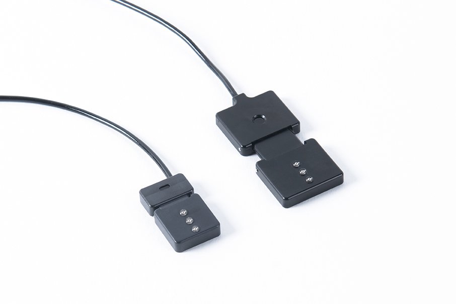
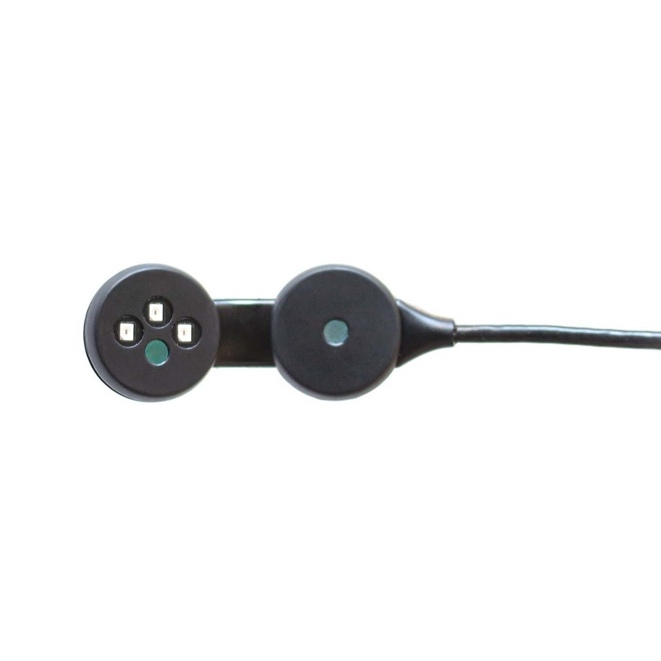
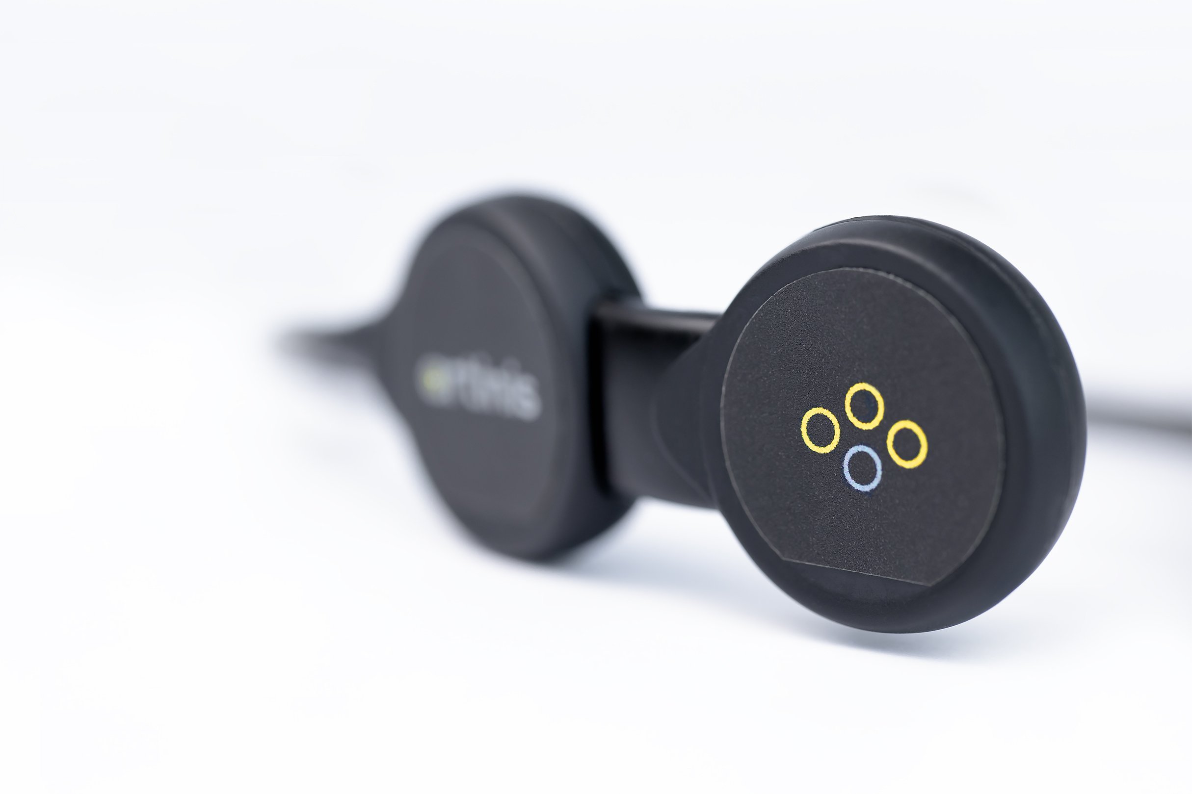
Figure 1: TSI is calculated using SRS with three transmitter (yellow) and one receiver (blue) optode
TSI - Tips and Tricks
The added value of measuring TSI is that it is an absolute measure, in contrast to oxygenated and deoxygenated hemoglobin concentration changes. Even though it is an absolute value, changes and baseline TSI values can differ between subjects due to differences in body composition, whereas TSI values are reproducible within subject. It is recommended to include a baseline (rest or continuous level of exercise) of at least 30 seconds before each intervention in your study design. An increase in body composition variability between subjects in a group will lead to an increase in the group TSI change variability.
Typical TSI values for measurements on human tissue are 55-80% during rest and if the subject is in a healthy condition. During exercise, the TSI in muscle tissue can drop to 10-20%. The TSI measured on the forehead will be relatively stable in many circumstances, but e.g. exercise in hypoxic conditions can cause a drop of 10% in TSI [3]. In the case of clinical conditions or extreme circumstances a larger drop in TSI is expected.
TSI Fit Factor
The Tissue Saturation Index (TSI) fit factor is an indicator of the TSI measurement quality. In SRS theory it is assumed that the tissue is homogeneous. However, tissue is never perfectly homogeneous. Small variations in tissue composition do not have an impact on the TSI measurement because hemoglobin is the main absorber of near-infrared light. Larger differences in regional tissue composition might impact the amount of light absorbed by the tissue and consequently influence the TSI measurement.
To help you achieve high-quality TSI measurements, we have developed the TSI fit factor algorithm. This algorithm compares the absorption in the tissue over multiple distances, in case of our devices three. When absorption levels are very similar, the TSI fit factor will be close to 100 (%). Typical values for the TSI fit factor are hard to provide since it is dependent on the tissue composition beneath the sensor. If you see a lower TSI fit factor than normal, make sure you did not place the device over birth marks, larger veins, hair or other larger irregularities. In case of large changes in the tissue during e.g. (heavy) exercise, the TSI fit factor might drop. Perfusion changes in the skin beneath the sensor might have a minor influence on the TSI measurement. The SRS algorithm uses the absorption differences over the different distances and therefore will compensate for homogeneous perfusion changes in the skin. Larger perfusion changes by e.g. bending the head or changing the position of body parts might increase the blood flow in the head and might impact the TSI. When interested in measuring TSI in brain tissue, related to task of interest, then blood flow due to gravity might be a confound.
NIRS devices that measures TSI
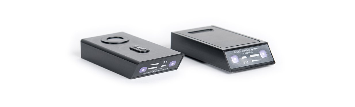
The PortaMon, PortaLite and PortaLite MKII devices all measure the TSI and provide the TSI fit factor, using three transmitters at different distances from the receiver. The devices are calibrated at delivery and should be recalibrated yearly. The devices do not need a warm-up time if placed on a human body.
Suggested readings:
[1] Fatigue following COVID-19 infection is not associated with autonomic dysfunction. Townsend L, Moloney D, Finucane C, McCarthy K, Bergin C, Bannan C, Kenny RA (2021).
[2] Cerebral hemodynamic response to a therapeutic bed for procedural pain management in preterm infants in the NICU: a randomized controlled trial. Ranger M, Albert A, MacLean K, Holsti L (2021).
[3] Changes in prefrontal cerebral oxygenation and microvascular blood volume in hypoxia and possible association with acute mountain sickness. Manferdelli G, Marzorati M, Easton C, Porcelli S (2021).
[4] Sensitivity and reliability of cerebral oxygenation responses to postural changes measured with near-infrared spectroscopy. Mol A, Woltering JHH, Colier WNJM, Maier AB, Meskers CGM, van Wezel RJA (2019).
[5] Reproducibility of NIRS assessment of muscle oxidative capacity in smokers with and without COPD. Adami A, Cao R, Porszasz J, Casaburi R, Rossiter HB (2017).
[6] The use of portable NIRS to measure muscle oxygenation and haemodynamics during a repeated sprint running test. Jones B, Hesford CM, Cooper CE (2013).


Functional Near-Infrared Spectroscopy is a neuroimaging modality measuring brain activity, that offers various advantages, such as easy usability and non-invasiveness. Thus, it can perfectly be applied to sensitive subjects, such as infants, toddlers, and the elderly. Read in this blog post, what makes fNIRS a suitable technology to measure neural activity in sensitive subjects, and how it is currently used in this population.