
Integrating muscle NIRS & HD-EMG: Gain advanced muscle insights
NIRS enables getting insights into local muscle oxygenation, whereas HD-EMG allows for receiving fast temporal and spatial information on muscle activity. Combining both portable and lightweight methodologies can provide significant advantages to further understand muscle activity and metabolism in many applications. Read this blog post to learn more about NIRS and HD-EMG, and what benefits simultaneous measurements of both can bring to your research.


What is Near-Infrared Spectroscopy (NIRS)?
Near-Infrared Spectroscopy (NIRS) measures relative concentration changes in oxygenated and deoxygenated hemoglobin in local (muscle) tissue using Near-Infrared Light. This can give information on local oxygenation and metabolism of any muscle. If you would like to learn more about the theory of using NIRS for muscle measurements, we recommend that you watch our webinar, Introduction to NIRS muscle measurements.
NIRS ApplicationsMost NIRS devices, such as our PortaMon MKIII, are completely portable, lightweight weight and easy to use. Hence, NIRS is used in a huge range of applications to measure muscle oxygenation, for instance, in sports sciences, exercise performance, clinical and rehabilitation studies, and hypoxia research.
What is High-Density Electromyography (HD-EMG)?


High-Density EMG (HD-EMG), also called high-density surface EMG (HDsEMG), measures spatial and temporal patterns of muscle activity using EMG signals recorded on the surface of the skin from at least four electrodes with close inter-electrode distances. The signal originates from the electrical activity of motor units in the skeletal muscle comprising a motor neuron and its innervated muscle fibers.
Benefits of HD-EMG over Bipolar EMG
Higher spatial resolution: Compared to Biopolar EMG, HD-EMG provides an increased number of electrodes placed close to each other. This enables measuring muscle activity more accurately and from a larger area of the muscle.
Accurate image reconstruction and muscle activity localization: The smaller distance between electrodes enhances spatial resolution, enabling advanced signal processing techniques such as decomposition and spatial filtering. This allows precise mapping of motor units and the exact location of muscle activation. HD-EMG can isolate the activity of individual motor units and track their firing patterns.
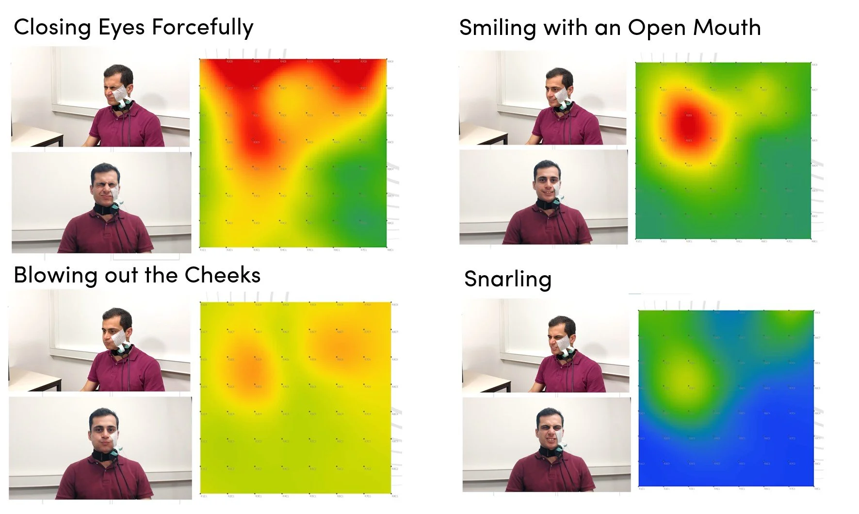
Figure 1: Heatmaps created during four different facial expressions using HD-EMG
HD-EMG ApplicationsHD-EMG systems such as the SPIRE are often portable and lightweight, but still enable accurate and precise measurements of muscle activity and localization. This allows for investigation of muscle fiber properties and for performing spatial muscle mapping (see Figure 1) or motor unit decomposition. Therefore, HD-EMG is applied in a huge range of application areas, including sports science, robotics, clinical and rehabilitation studies, and body mechanics.
Would you like to learn more about the creation of spatial muscle mapping with our HD-EMG devices? Then watch this video on a Signal Demo: Facial Muscles HD-EMG Recording.
Why Combining NIRS and HD-EMG?
Combining mNIRS and HD-EMG can help to get a complete picture on functional, neuromuscular and metabolic state of a muscle. Both techniques are portable, light weight and relatively easy to use, which enables provision of real-time feedback in dynamic studies involving movement and in every setting.
When assessing fatigue mechanisms, HD-EMG can determine changes in motor unit recruitment, firing rates and conduction velocity during exercise. NIRS on the other hand can detect oxygen desaturation and recovery dynamics. Combining both provides a comprehensive view of fatigue on the neural (central) and metabolic (peripheral) components.
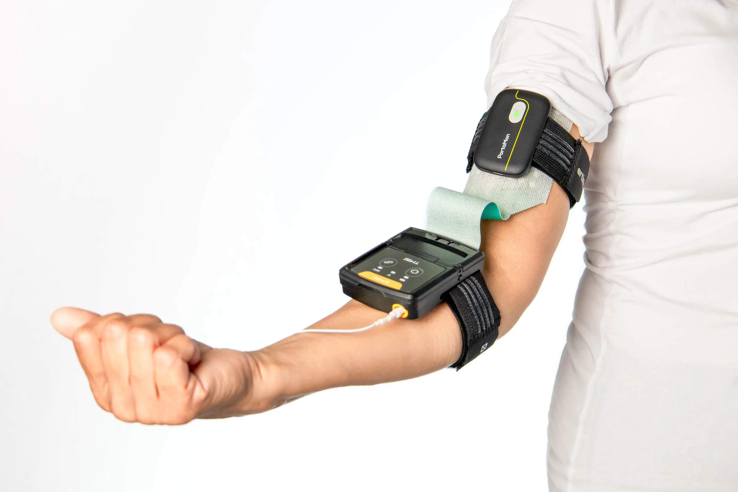
Additionally, combining detailed activation patterns with HD-EMG, and local changes in muscle oxygenation measured with NIRS, a full spatial picture on muscle activation and their response to metabolic demands can be achieved. Additionally, our NIRS - HD-EMG combinations, such as PortaMon and SPIRE, make it easy to integrate both technologies within a single measurement setup.
Further, combining HD-EMG and NIRS can help to get complete physiological insights in effectiveness of training and rehabilitation strategies. By simultaneously monitoring changes in muscle activity with HD-EMG and oxygen utilization with NIRS before, during and after intervention can help to 1. identify whether performance or recovery is limited by neural drive or by oxygen delivery and 2. understand whether changes or improvements over time come from better oxygen support or from more efficient recruitment strategies.
Would you like to learn more about our portable HD-EMG & NIRS devices and how they can be combined for muscle research? Then feel free to reach out at askforinfo@artinis.com!
Suggested Literature
Bhambhani Y, Fan JL, Place N, Rodriguez-Falces J, Kayser B. Electromyographic, cerebral, and muscle hemodynamic responses during intermittent, isometric contractions of the biceps brachii at three submaximal intensities. Front Physiol. 2014 Jun 11;5:190.
Di Giminiani R, Cardinale M, Ferrari M, Quaresima V. Validation of Fabric-Based Thigh-Wearable EMG Sensors and Oximetry for Monitoring Quadriceps Activity during Strength and Endurance Exercises. Sensors (Basel). 2020 Aug 19;20(17):4664.
Felici F, Quaresima V, Fattorini L, Sbriccoli P, Filligoi GC, Ferrari M. Biceps brachii myoelectric and oxygenation changes during static and sinusoidal isometric exercises. J Electromyogr Kinesiol. 2009 Apr;19(2):e1-11.
Guo W, Sheng X, Liu H, Zhu H. Toward an Enhanced Human–Machine Interface for Upper-Limb Prosthesis Control With Combined EMG and NIRS Signals. IEEE Transactions on Human-Machine Systems. Vol. 47, no. 4, pp. 564-575, Aug. 2017.
Kauppi K, Korhonen V, Hany F, Kallio M, Myllyiä T. Combined surface electromyography, near-infraredspectroscopy and acceleration recordings of musclecontraction: The effect of motion. Journal of Innovative Optical Health Sciences. Vol. 10, No. 2 (2017) 1650056.
Scano A, Zanoletti M, Pirovano I, Spinelli L, Contini D, Torricelli A, Re R. NIRS-EMG for Clinical Applications: A Systematic Review. Applied Sciences. 2019; 9(15):2952.


Both NIRS and HD-EMG are non-invasive and portable techniques for muscle measurements. Combining these two methodologies can provide complementary information and enable enhanced insights into muscle activity and metabolism in many applications. In this blogpost we explain both techniques and highlight the advantages of combining NIRS and HD-EMG!