Near infrared spectroscopy (NIRS) relies on two characteristics of human tissue. The relative transparency of tissue to light in the NIR range and the oxygenation-dependent light-absorbing characteristics of hemoglobin.
The theory of Near Infrared Spectroscopy
What is Near Infrared Spectroscopy?

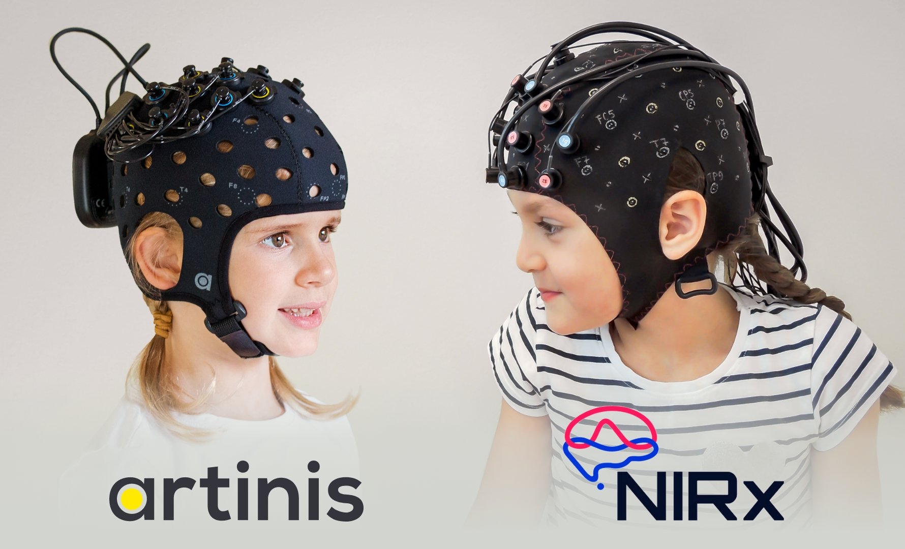
Near infrared spectroscopy (NIRS), the technique on which Artinis devices e.g. The OxyMon, Brite family, and PortaMon are based, relies mainly on two characteristics of human tissue. First, the relative transparency of tissue to light in the NIR range, and second, the oxygenation-dependent light absorbing characteristics of hemoglobin.
By using a number of different wavelengths, the relative changes in hemoglobin concentration can be displayed continuously.
Using the NIRS principle, it becomes possible to monitor:
Non-invasively
Affordably
Continuously
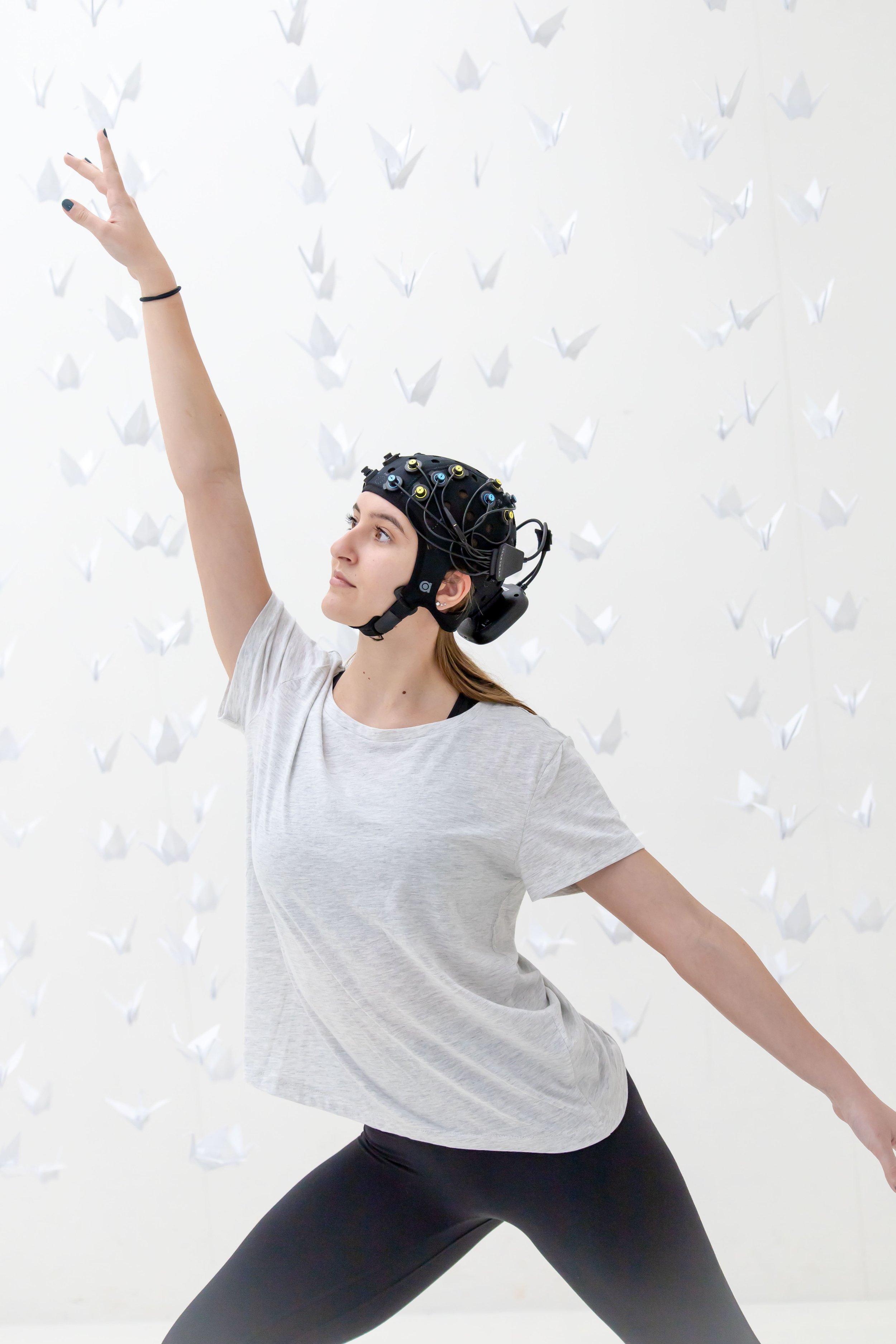
In the lab, or even in the field
Without the need for special infrastructure
Without specially trained personnel
Why NIRS technique?
All cells in all organs of the body have a constant but variable need for oxygen. However, the body stores for oxygen are minimal. A constant and adequate supply of oxygen to the tissues through the circulation is essential. Any interference with tissue oxygenation will lead very rapidly to irreversible damage.
Optical oximetry, and near infrared spectroscopy (NIRS) in particular, is a tool for assessing of the oxygenation status and hemodynamics of various organs, e.g. muscle and brain.
NIRS Explained by Artinis
What is NIRS or fNIRS and how does it work?
In this video series, each of our application specialists, active in the field, explains what you must know in the world of (functional) Near-Infrared Spectroscopy.
How Near Infrared Spectroscopy (NIRS) started
NIRS started with a paper published by Frans Jöbsis in Science (1977), Jöbsis reported that biological tissues are relatively transparent to light in the near infrared (700-1300 nm).
Therefore, it is possible to transmit enough photons through organs for in situ monitoring. In this near infrared region, hemoglobin - including its two main variants oxyhemoglobin (O2Hb) and deoxyhemoglobin (HHb)- exhibits oxygen-dependent absorption. Hemoglobin is assumed to be the main chromophore in biological tissue that absorbs light in this near infrared region.
The science
If the absorption is known, the Lambert-Beer law can be used to calculate the chromophore's absorption. The Lambert-Beer law is given by:

ODλ is a dimensionless factor known as the optical density of the medium, I0 is the incident light, I the transmitted light, ελ the chromophore's extinction coefficient (in mM-1•cm-1), c is the concentration (in mM) of the chromophore, L the distance (in cm) between light entry and exit points and λ is the wavelength used (in nm).
The Lambert-Beer law is intended to be used in a transparent, non-scattering medium. When it is applied to a scattering medium (Figure 1.1), e.g. biological tissue, a dimensionless pathlength correction factor must be incorporated. This factor, sometimes called the differential pathlength factor (DPF), accounts for the increase in optical pathlength due to scattering in the tissue. The modified Lambert-Beer law for a scattering medium is given by:

where ODλ represents the oxygen-independent optical losses due to scattering and absorption in the tissue. Assuming that ODλ is constant during a NIRS measurement, we can convert the change in optical density into a change in concentration.
This equation is valid for a medium with one chromophore. If more chromophores are involved, we need to measure at least as many wavelengths as there are chromophores present. This results in a set of linear equations. The solution of this set leads to the algorithm used in most NIRS systems. A scattering medium makes it possible to measure the absorption with the near infrared source and detector parallel to each other (Figure 1.1). This offers the opportunity to measure oxygenation in larger tissues, e.g. muscles and brain using NIRS equipment.

A scattering medium makes it possible to measure the absorption with the near infrared source and detector parallel to each other (Figure 1.2). This offers the opportunity to measure oxygenation in larger tissues, e.g. muscles and brain using NIRS equipment.
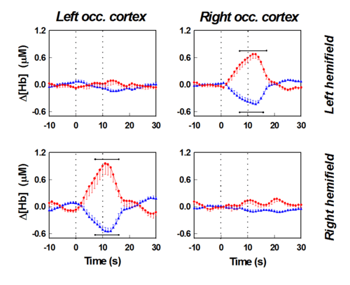
Visual Cortex Stimulation
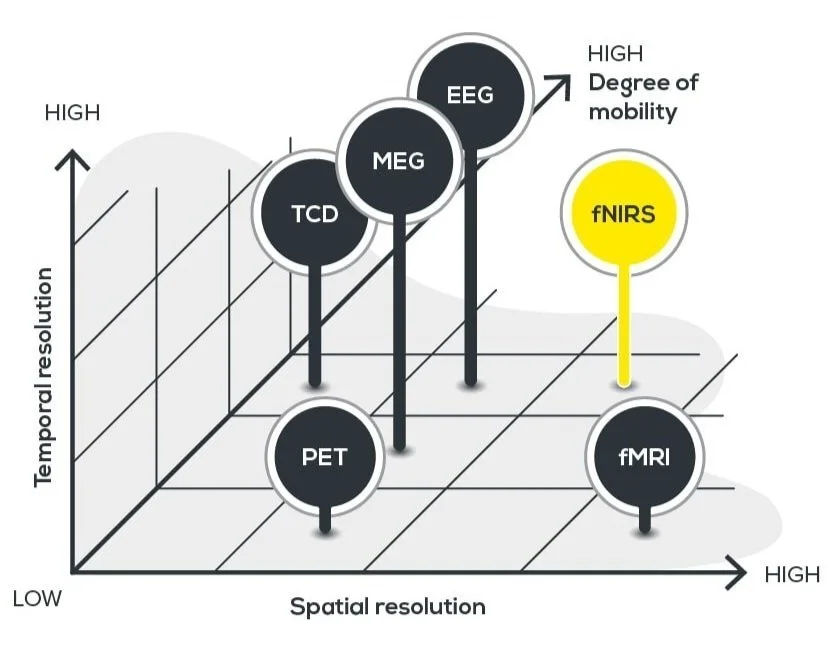
NIRS compared to other modalities.
NIRS algorithm
Defining the algorithm used by NIRS requires the spectral extinction coefficients of the various chromophores. The spectra of the two main chromophores, O2Hb and HHb.
The sum of O2Hb and HHb is a measure of the total blood volume (tHb) in the tissue. Muscle tissue contains two further chromophores: oxy- and deoxymyoglobin (O2Mb and HMb). In order to distinguish between hemoglobin and myoglobin in muscle tissue, the spectra need to be sufficiently different. Unfortunately, this is not the case in the near-infrared region of the spectrum. This means, NIRS cannot distinguish if the measured oxygen concentration is carried by hemoglobin or myoglobin. The wavelengths that can distinguish the Hb and Mb are not able to penetrate the tissue deep enough.
What’s the difference between NIRS and pulse oximetry?
The technique on which near-infrared spectroscopy relies is closely analogous to the technique of pulse oximetry.
The main difference is the tissue being sampled. Pulse oximetry calculates the percentage of oxygenated hemoglobin in the arterial blood. NIRS calculates the changes in oxy- and deoxyhemoglobin (and optionally the percentage of oxygenated hemoglobin) in the tissue under investigation (capillaries), which contains both arterial and venous blood.
(f)NIRS introduction course
Live INTRODUCTION TO (FUNCTIONAL) NEAR-INFRARED SPECTROSCOPYWith so much to explore in the (f)NIRS world, learning the fundamentals from an expert can be incredibly valuable. That's why we offer periodic in-person/online (f)NIRS introduction courses designed to help you grasp both the fundamental science and hands-on use of the devices.
How can we help you?
(f)NIRS applications
For your convenience, we have compiled a list of all (f)NIRS-publications performed with our equipment. Additionally, you can also find an overview of the latest (f)NIRS publications on this page as well.
How does (f)NIRS device work?
Are you interested in trying out our (f)NIRS devices, but the conferences are out of reach?
We are coming to you with the possibility of a 30-minute online demo!
Follow us
Subscribe to Artinis on YouTube or other social media channels to stay informed about the latest research, technology, and relevant events regarding (f)NIRS.