
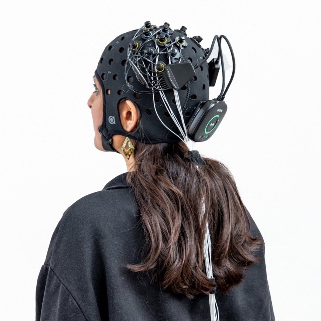
Introduction to multimodal fNIRS — EEG measurements
This is the first post and the introduction to our blog series on Artinis solutions for combined functional near-infrared spectroscopy (fNIRS) and electroencephalographic (EEG) measurements.
We know that it can seem difficult to integrate a new technique into your experimental set up, and to navigate the possibilities that two or more techniques offer together. With this blogpost series, we aim to facilitate this process for you. In several posts, we will present the potential of combined fNIRS-EEG research and introduce how this can be easily achieved with our user-friendly products.
First of all, it is worth mentioning that recent advancements in neuroimaging techniques have made it easier and easier to investigate the functionality of the brain. Yet, each of these techniques measures specific neurophysiological mechanisms, operating at different temporal and spatial scales, and therefore individually none of them provides a full picture of brain dynamics.
Fortunately, it is possible to perform multimodal simultaneous recordings of brain signals. That is, one can use several technologies at the same time and combine the information that each of them provides. One such combination that has gained a lot of interest in the last few years is, in fact, fNIRS+EEG.
The hallmarks of fNIRS and EEG as brain measuring techniques
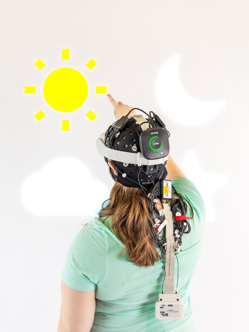
fNIRS is a fairly new technique that uses near infrared light to quantify changes in concentration of oxy- and deoxyhemoglobin in the brain. More specifically, fNIRS can measure the hemodynamic response that occurs in the brain when an area becomes more active and demands more energy. This response is relatively slow; it takes place 5 to 10 seconds after stimulus appearance, but it can be easily localized with fNIRS (good spatial resolution).
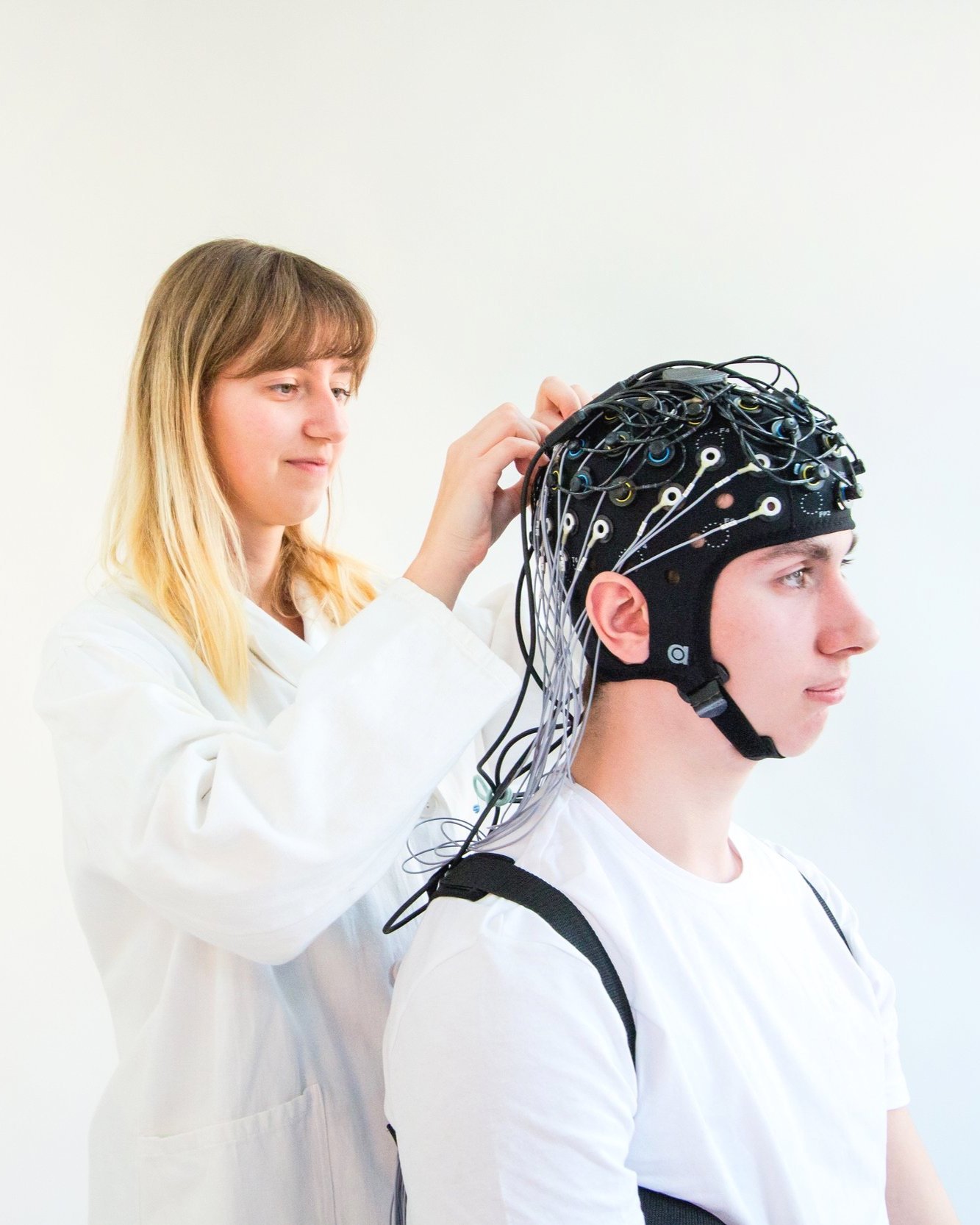
In contrast, EEG is a technique that has been around for more than a century, and is therefore already for many years widely used to measure brain activity. In particular, EEG captures the electrical activity of the brain, on a macroscopic level, by placing electrodes on the head. Contrary to the hemodynamic response, brain electrical activity is fast paced, and can be measured on a scale of milliseconds. However, since the electrodes are placed on the scalp, volume conduction plays a part in EEG, and activity is not as easily localized. Therefore, EEG has higher temporal resolution, but lower spatial resolution than fNIRS.
The benefits of combining fNIRS and EEG

Figure 1: Comparison of fNIRS and other neuroimaging technologies
As outlined above, fNIRS and EEG provide different physiological information about brain cortex activity (i.e., brain electrical and hemodynamic-metabolic activity). This means that the information provided by both techniques can complement each other and therefore, together they offer a more complete picture.
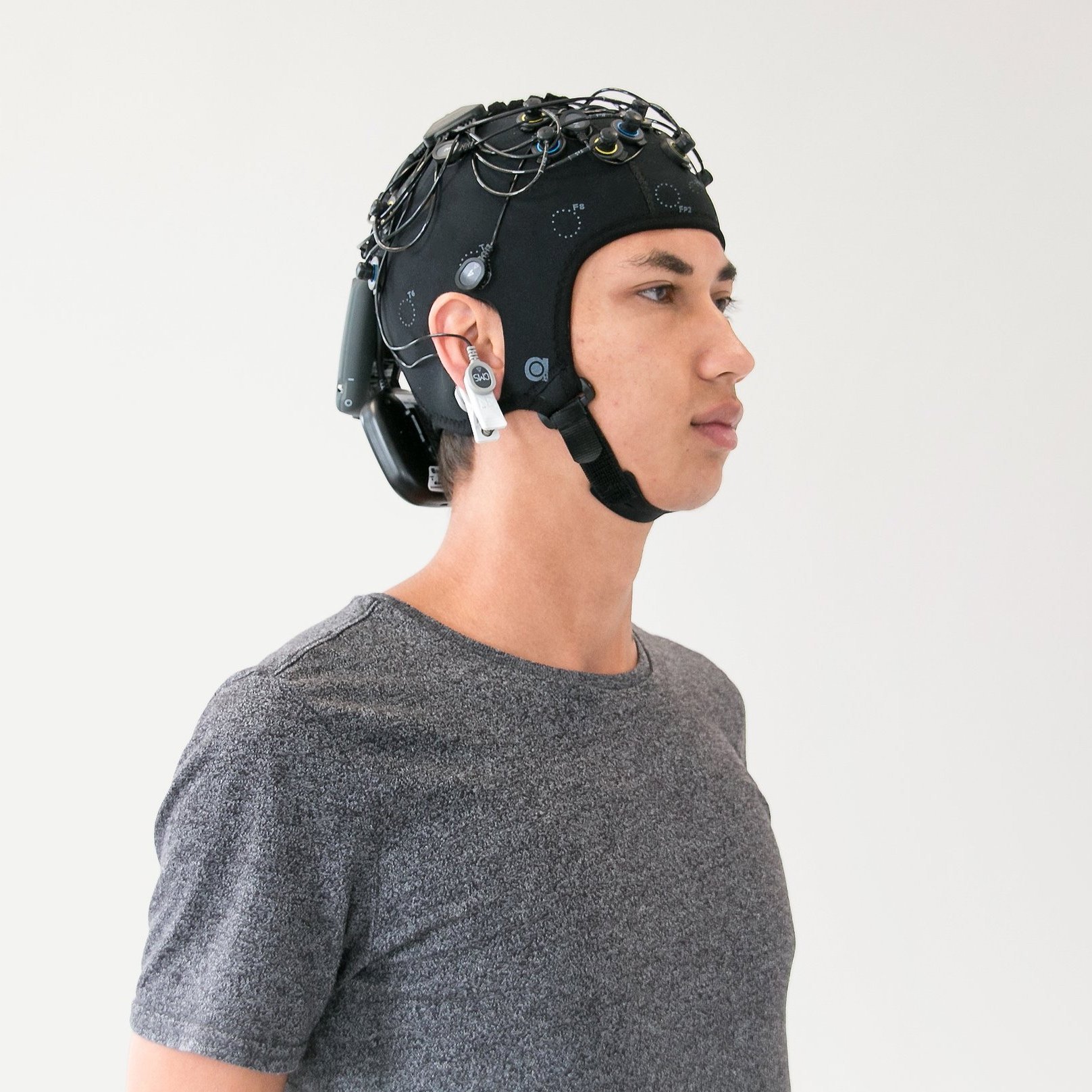
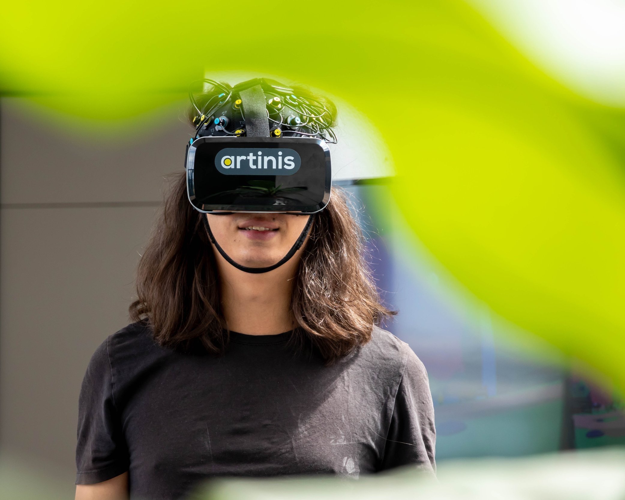
In addition, both technologies are non-invasive, as well as relatively affordable and mobile (wearable). In comparison to other neuroimaging techniques, such as fMRI, PET, MEG…, combined EEG-fNIRS mobility also allows for a larger variety of experimental set-ups, including more naturalistic studies. For the same reasons, this combination is very suitable for a wide range of populations, from elderly to newborns and from healthy participants to patients with different medical conditions.
The challenges of combining fNIRS and EEG
Researchers have had to face various challenges when combining fNIRS and EEG.
First, since usually two different devices are used for data collection, an extra step is required to synchronize both data streams.
Second, due to the substantial difference between the time scales at which fNIRS and EEG operate, it can be difficult to think of a suitable experimental design that works for both.
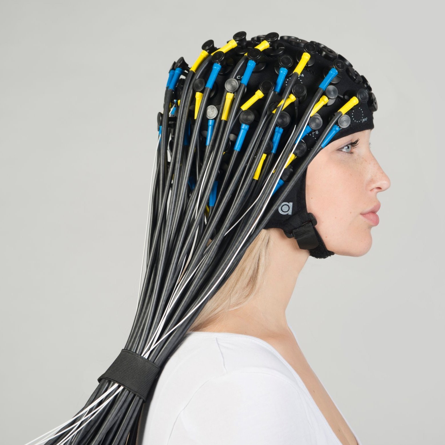
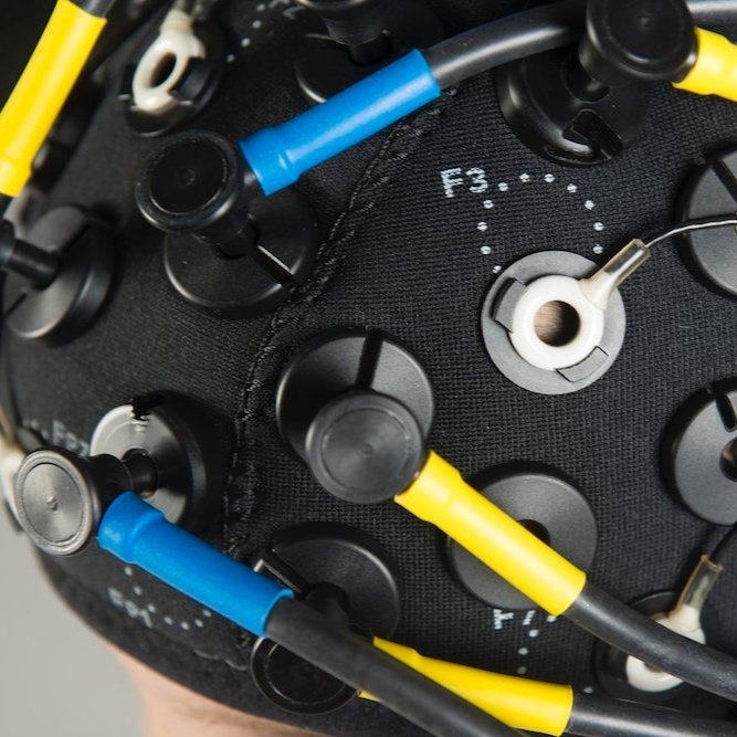
Third, both technologies make use of the scalp to place the sensors (electrodes and optodes). Thus, the available surface for this had to be shared between the two, decreasing the number of each type of sensor that can be utilized, and consequently decreasing the number of possible EEG and fNIRS measuring channels.
Finally, fNIRS optodes often contain electronic parts, which can interfere with the EEG signal, adding noise to it.
Talk to us about your fNIRS - EEG setup!
We are happy to think along with you if you tell us about your research at askforinfo@artinis.com
In the following blog posts of this series, we will discuss how we at Artinis have tackled these challenges individually to take the integration of fNIRS and EEG to the next level.


When simultaneously measuring fNIRS and EEG, placement of both devices should ideally ensure proper coverage of the desired measurement location, minimize interference and take into account (technical) characteristics and basic of both techniques. Read this blog post to learn more about relevance of these points and further recommendations for integrating fNIRS and EEG on one head.